Challenge: A Complex Bone Deformity Requiring Precision Planning
Eighteen-year-old Moises Campos faced a significant orthopedic challenge. A tibial tubercle fracture from a previous injury had caused a growth arrest below his kneecap, leading to a deformity in his tibia. This condition affected his ability to walk and run and put him at risk for early-onset arthritis.
To correct the deformity, Dr. Afshin Aminian, Medical Director of the Children’s Hospital of Orange County (CHOC) Orthopedic Institute, determined that the best approach was to cut the bone and slowly reposition it. However, the complex nature of Moises’ deformity made intraoperative assessment difficult. A traditional CT scan alone would not provide the full spatial understanding necessary for precise surgical execution.
Dr. Aminian and his surgical team sought a solution that would allow them to visualize, study, and plan the procedure in greater detail before stepping into the operating room.
Solution: Patient-Specific 3D Printed Bone Models
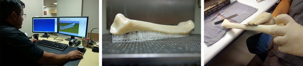
To improve surgical precision, Dr. Aminian partnered with Dinsmore Inc., a 3D printing and additive manufacturing expert in Orange County, to create an accurate, life-size 3D model of Moises’ tibia.
Using CT scan data provided by CHOC, Dinsmore’s in-house designers:
- Converted the anatomical data into a high-fidelity surface model.
- 3D printed multiple versions of the tibia using stereolithography (SLA) technology, known for its precision and speed.
- Refined the post-processing to ensure the density and surface texture resembled real bone, allowing for realistic surgical practice.
“The front-end work of ensuring data integrity was the most time-intensive step, but also the most important. At Dinsmore, we take pride in offering our customers design for prototyping to ensure that their files are optimized for successful prints.”
— Jay Dinsmore, Founder & CEO, Dinsmore Inc.
Results: Improved Surgical Planning & Patient Outcomes
- With the 3D printed bone model in hand, Dr. Aminian’s team was able to:
- Plan precise surgical cuts and fit the bone with the correct external fixation construct before surgery.
- Reduce intraoperative guesswork, leading to a smoother and more efficient procedure.
- Improve patient and family education by providing a tangible model to explain the surgical plan.
“For complex surgeries like this, the 3D model was an invaluable tool for pre-surgical planning. You can measure deformities in CT scans, but there’s nothing like holding the physical model in your hands.” — Dr. Justin Roth, Pediatric Surgeon, CHOC
Outcome: Faster Recovery & Restored Mobility
Thanks to the detailed pre-surgical planning enabled by Dinsmore’s 3D printing expertise, Moises underwent a successful surgery that restored his normal gait and function within just four months. Within six months, he had returned to his active lifestyle without restrictions.
His stepmother expressed her gratitude for the life-changing impact of the surgery, stating:
“The surgery has had a genuine impact on his life. He is more confident, engages more with others, and is excited to participate in activities he used to avoid. We are incredibly thankful for the surgical team at CHOC.”
Conclusion: Advancing Pediatric Surgery Through Additive Manufacturing
By leveraging patient-specific 3D printing, CHOC surgeons and Dinsmore Inc. demonstrated how advanced technology can improve surgical precision, reduce risks, and enhance patient outcomes.
This collaboration highlights the transformative role of additive manufacturing in modern medicine—bringing innovation to life, one patient at a time.